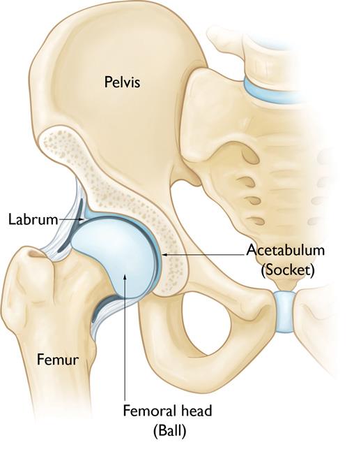
The hip is a ball-and-socket joint. The socket is formed by the acetabulum, which is part of the large pelvis bone. The ball is the femoral head, which is the upper end of the femur (thighbone).
A slippery tissue called articular cartilage covers the surface of the ball and the socket. It creates a smooth, frictionless surface that helps the bones glide easily across each other.

The acetabulum is ringed by strong fibrocartilage called the labrum. The labrum forms a gasket around the socket
The joint is surrounded by bands of tissue called ligaments. They form a capsule that holds the joint together. The undersurface of the capsule is lined by a thin membrane called the synovium. It produces synovial fluid that lubricates the hip joint.
Your doctor may recommend hip arthroscopy if you have a painful condition that does not respond to nonsurgical treatment. Nonsurgical treatment includes rest, physical therapy, and medications or injections that can reduce inflammation.
Hip arthroscopy may relieve painful symptoms of many problems that damage the labrum, articular cartilage, or other soft tissues surrounding the joint. Although this damage can result from an injury, other orthopaedic conditions can lead to these problems, including:
Your orthopaedic surgeon may recommend that you see your primary doctor to assess your general health before your surgery. He or she will identify any problems that may interfere with the procedure. If you have certain health risks, a more extensive evaluation may be necessary before your surgery.
If you are generally healthy, your hip arthroscopy will most likely be performed as an outpatient. This means you will not need to stay overnight at the hospital.
Be sure to inform your orthopaedic surgeon of any medications or supplements that you take. You may need to stop taking some of these before surgery.
The hospital or surgery center will contact you ahead of time to provide specific details of your procedure. Make sure to follow the instructions on when to arrive and especially on when to stop eating or drinking prior to your procedure.
Before the operation, you will also be evaluated by a member of the anesthesia team. Hip arthroscopy is most commonly performed under general anesthesia, where you go to sleep for the operation. Regional anesthesia, such as spinal or epidural, can also be used. With regional anesthesia, you are awake but your body is numb from the waist down. Your orthopaedic surgeon and your anesthesiologist will talk to you about which method is best for you.
Traditional hip replacement prostheses only allow a fairly narrow range of shapes and sizes. Even small changes in hip geometry can have large effects on the function of the hip joint. New types of hip replacement are now available that allow a far wider range of sizes and lengths. New pre-operative 3-D imaging
Hip arthroscopy means keyhole surgery of the hip joint. Under an anaesthetic, a camera on the end of a telescope can be passed into the hip joint. The inside of the hip can be visualised, helping confirm a diagnosis. Damage inside the hip, such as loose bits of cartilage or cartilage tears can be treated.
Hip arthritis causes pain, stiffness and difficulty walking. The pain can be felt deep in the hip, in the groin, in the thigh or even in the knee. Early arthritis can be helped by anti-inflammatories, physiotherapy or injections into the joint. Severe arthritis requires hip replacement surgery.
Hip resurfacing is one specific type of hip replacement. With hip resurfacing, instead of the ball part of the joint being removed and replaced, it is shaved and then resurfaced with a circular metal cap. The choice of whether to have a traditional hip replacement or a hip resurfacing procedure is complex, and should be discussed with your surgeon. The risks of the surgery are pretty much the
In patients with hip pain, a diagnostic injection of anaesthetic into the joint can help clarify the diagnosis, in terms of working out exactly where the pain comes from. Injection of cortisone / steroid or hyaluron can give relief of symptoms from hip arthritis. In some cases where patients either have suspected or proven pathology inside the hip joint, intra-articular injection of the hip may well prove beneficial.
The hip socket is called the ‘acetabulum’. Around the rim of the socket there is a little lip of cartilage – the ‘labrum’. An acetabular labral tear is where this lip of cartilage around the rim tears. It causes sharp catching pains in the hip – often felt in the groin or thigh. If symptoms fail to settle with rest / physiotherapy then you need to see a specialist surgeon. The best investigation is a 3T (a high-res 3-Tesla) MRI scan. Treatment can be rehabilitation or hip arthroscopy (keyhole surgery of the hip).
The hip joint is a ball and socket joint. There is a layer of cartilage covering the ball and a layer of cartilage lining the socket. Hip arthritis is where this cartilage wears away, eventually exposing bare bone. Arthritis in the hip causes pain and stiffness. The pain can be felt in the hip, groin, thigh or even in the knee. Early arthritis may respond to painkillers, physiotherapy, exercise or hip injections. Advanced arthritis is treated by hip replacement surgery.
There is a little sac of fluid on the outer side of the hip, called a bursa. It overlies the lump of bone on the outer side of the hip called the greater trochanter. The sac is therefore called the ‘trochanteric bursa’. This sac can become inflamed – this is called ‘trochanteric bursitis’. This causes pain and tenderness on the outer side of the hip and thigh. Patients often find it painful to walk or to lie on the affected side in bed. The investigation of choice is either an ultrasound scan or an MRI scan.
Hip impingement is also called ‘femoroacetabular impingement’. It occurs when the edge of the ball of the hip joint rubs against the edge of the socket. It causes pain in the hip, groin, thigh or even knee. There may be sudden sharp pains or catching sensations. If symptoms fail to settle with rest or physiotherapy then you need to see a surgeon. An MRI scan with dye injected into the hip may be required. Treatment can be performed with a hip arthroscopy (keyhole surgery of the hip) .

5000

1000

10000

750